40 microscope parts and labels
› Microscope-ANNLOV-ElectronicAmazon.com : LCD Digital Microscope,ANNLOV 4.3 inch Handheld ... Feb 05, 2020 · This item LCD Digital Microscope,ANNLOV 4.3 inch Handheld USB Microscope 50X-1000X Magnification Coin Microscope Video Camera with 8 Adjustable LED Lights for Adults PCB Soldering Kids Outside Use ANNLOV 7" LCD Digital Microscope with 32GB TF Card 1200X Maginfication 1080P Coin Microscope with Wired Remote,12MP Ultra-Precise Focusing Video ... Amazon.com : LCD Digital Microscope,ANNLOV 4.3 inch … Web05.02.2020 · This item LCD Digital Microscope,ANNLOV 4.3 inch Handheld USB Microscope 50X-1000X Magnification Coin Microscope Video Camera with 8 Adjustable LED Lights for Adults PCB Soldering Kids Outside Use ANNLOV 7" LCD Digital Microscope with 32GB TF Card 1200X Maginfication 1080P Coin Microscope with Wired …
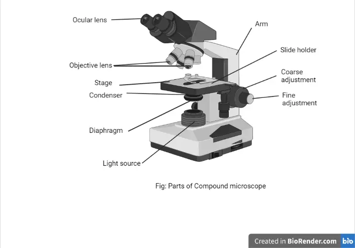
15 Microscope Parts with Diagram, Location and Function - Study Read But a compound microscope has many parts like Eyepiece Body Tube Revolving nosepiece Objective lens (Three 10x, 45x, 100x) Coarse adjustment Fine adjustment Arm/handle Rack stop Fixed stage Mechanical Stage Clips Side-ward movement knob Front and back movement knob Condenser and Diaphragm Mirror (convex and concave mirror) Base Pillars

Microscope parts and labels
Barcode Labels and Tags | Zebra WebWith more than 400 stocked ZipShip paper and synthetic labels and tags – all ready to ship within 24 hours – Zebra has the right label and tag on hand for your application. From synthetic materials to basic paper solutions, custom to compliance requirements, hard-to-label surfaces to easy-to-remove labels, or tamper-evident to tear-proof, we have more … Microscopy- History, Classification, Terms, Diagram - The Biology Notes History of Microscope. In the 1 st Century AD, the Romans invented the glass and used them to magnify objects. In the early 14 th Century AD, eyeglasses were made by Italian spectacle makers. In 1590, two Dutch spectacle makers, Hans, and Zacharias Jansen created the first microscope. It was a simple tube with 2 lenses system and had 9X ... Compound Microscope - Types, Parts, Diagram, Functions and Uses It comes with a wide body and base. Its distinct parts include a condenser, illumination, focus lock, mechanical stage, and a revolving nosepiece which can hold up to five objectives. It usually has a binocular head, which makes long-term observation easy. Image 22: An example of a research compound microscope.
Microscope parts and labels. Simple Microscope - Diagram (Parts labelled), Principle, Formula and Uses simple microscope has mainly two types of parts - Optical and Mechanical. The optical parts include - Lens - Mirror - Eyepiece The mechanical parts include - Arm - Stage - Nosepiece - Base - Coarse focusing mechanism - Fine focusing mechanism Q 5. What are the three types of microscopes? The three types of microscopes are Microscope, Microscope Parts, Labeled Diagram, and Functions The description given below summarize the brief description of microscope parts used to visualize the microscopic specimens such as animal cells, plant cells, microbes, bacteria, viruses, microorganisms etc. The Microscopes parts divided into three different structural parts Head, Base, and Arms. Stereo Microscope - Parts, Types and Uses - Laboratoryinfo.com Uses. A stereo microscope is primarily used to view specimens like plants and animals. A stereo microscope is useful when working with circuits and watches. It can be used for microsurgery. It is used to view crystals. (2,3, 4, and 5) Image 2: The different parts of a stereo microscope. Picture Source: microbehunter.com. SMT Supplies WebSMT Supplies 21088 Bake Parkway Suite 102 Lake Forest, CA 92630 (888) 449-6655. contact@smtsupplies.com
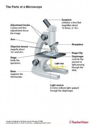
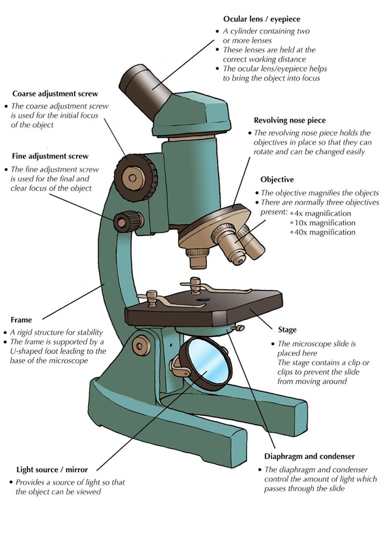
Compound Microscope- Definition, Labeled Diagram, Principle, Parts, Uses Parts of a Compound Microscope Eyepiece And Body Tube. The eyepiece is the lens through which the viewer looks to see the specimen. It usually contains a 10X or 15X power lens. The body tube connects the eyepiece to the objective lenses. Objectives and Stage Clips Objective Lenses are one of the most important parts of a Compound Microscope. All About Microscopes for Kids — with Free Printables Whatever microscope you choose, be sure to grab our free printable microscope activity set. This set includes parts of the microscope (labeled and with labels for the student to add), a word search, writing page and several sheets for drawing observations from under the microscope. You can grab the printables from our online shop. LAS X Industry Microscope software for Industry | Products WebMeasure parameters, such as the length, area, diameter, angle, or perimeter of objects you mark with adjustable tracing lines, drawing directly in the live images. Add labels for easy analysis. Apply measurements to several images to determine statistical trend and compare data in measurement templates. Apply long-distance measurements that ... Labeling a Microscope Free Worksheet Pack - Homeschool Giveaways Then, use the worksheet pack and this post to talk through all the parts of the microscope with your kids. As you discuss the function of each piece, have them label the parts of a microscope on the printable. Once they have the microscope parts labeled, see if they can use the microscope to look at an image of the specimen.
Microscope Parts Explained - GigOptix The main job of these parts is to support the instrument, so they will typically be the heaviest components of the microscope compared to the optical components. These parts will also house other parts of the microscope, such as lenses and knobs. Head/Body: One of the main microscope parts is the head, or otherwise known as the body. rsscience.com › stereo-microscopeParts of Stereo Microscope (Dissecting microscope) – labeled ... The objective lenses are the most important parts of a microscope. Compared to a compound microscope where the objectives attached to the nosepiece can be seen and identified individually (based on color bands and their respective labels), the objectives of a dissecting microscope are located in a cylindrical cone and, therefore, are not ... proscitech.com.auProSciTech Laboratory supplies and Lab equipment for Histology, Pathology, Light Microscopy, Electron Microscopy and specialist researchers. Parts of a microscope with functions and labeled diagram - Microbe Notes There are three structural parts of the microscope i.e. head, base, and arm. Head - This is also known as the body. It carries the optical parts in the upper part of the microscope. Base - It acts as microscopes support. It also carries microscopic illuminators.
Parts of a Microscope - Free Printable | Learning science ...
Microscope: Types of Microscope, Parts, Uses, Diagram - Embibe There microscope anatomy includes three structural parts, i.e. head, base, and arm. Head - This is also known as the body; it carries the optical parts in the upper part of the microscope.. Base - It acts as microscopes support.It also carries microscopic illuminators. Arms - The microscope arm connects the base and the head and the eyepiece tube to the microscope base.

Microscopy and Its Types - BIOLOGY EASE
Brightfield Microscope (Compound Light Microscope)- Definition ... The arm: This is a sturdy metallic backbone of the microscope, used to carry and move the microscope from one place to another. They also hold the microscope base which is the stand of the microscope. The arm and the base hold all the microscopic parts. It has a light illuminator or a mirror found at the base or on the microscope's nosepiece.

Compound Microscope- Definition, Labeled Diagram, Principle ...
Parts of Stereo Microscope (Dissecting microscope) – labeled … WebOptical parts of a stereo microscope work together to magnify and produce a 3-D image of the specimens. These parts include: Eyepieces. The eyepiece (or ocular lens) is the lens part at the top of a microscope that the viewer looks through. Typically, standard eyepieces for a dissecting microscope have a magnifying power of 10x. Optional eyepieces of …

Label the Microscope Diagram | Quizlet
› lesson-plans › scientific-methodScientific Method Worksheets - The Biology Corner Microscope Use. How to Use a Microscope – basic guidelines, tips and troubleshooting for the classroom light microscope | Presentation. Microscope Labeling – image, no labels Microscope Coloring – learn the parts of the microscope by coloring. Microscope “E” Lab – use a microscope to examine the letter “e”

Compound Microscope Parts, Functions, and Labeled Diagram ...
› Magnifying-Magnifier-HeadbandAmazon.com: Magnifying Glasses 8X 15X 23X Magnifier LED ... About this item . Double eye magnifying glasses magnifier loupe, with 2pcs adjustable LEDs to help it work in low-light conditions ; Left right double eye patches magnifierloupe with adjustable LED to help work in low-light conditions.Set of 2 magnifying glasses mounted on a one-size-fits-all eyeglass frame for easy hands-free operation.

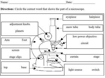
13 - Microscope Parts - PowerPoint Worksheet.docx - 1 Name: _ ...
Bright-field microscope (Compound light microscope) - Diagram (Parts ... Bright-field microscope parts (Labeled Diagram) Ocular Lens This microscope has two eye lenses or ocular lens on the top of the microscope that are used to focus the image from the objective lens. It is from these lenses that we see the magnified image of the specimen. Objective Lens

The Microscope
Free Microscope Worksheets for Simple Science Fun for Your Students WebParts of a Microscope . The first worksheet labels the different parts of a microscope, including the base, slide holder, and condenser. If you have a microscope, compare and contrast this worksheet to it. Also, your kids can color this microscope diagram in and read the words to each part of the microscope. Then, you can have your children study the …
The Parts of a Microscope (Labeled) Printable Printable (6th ...
Parts of a Microscope: Lesson for Kids - Study.com Sometimes when you look it's very dark. Slide your finger down to the base of the microscope and look for a wheel that you can turn. This controls the light intensity coming through the image. Turn...

Microscope Parts & Functions - AmScope
Types and parts of microscopes | Kenhub This discussion will cover the general anatomy of light and electron microscopes, their parts, the different subtypes of each, as well as the advantages and disadvantages of each. Additionally, it will also look at how each of these devices has helped in the advancement of the field of medicine. Contents Light microscopy Parts Visualisation
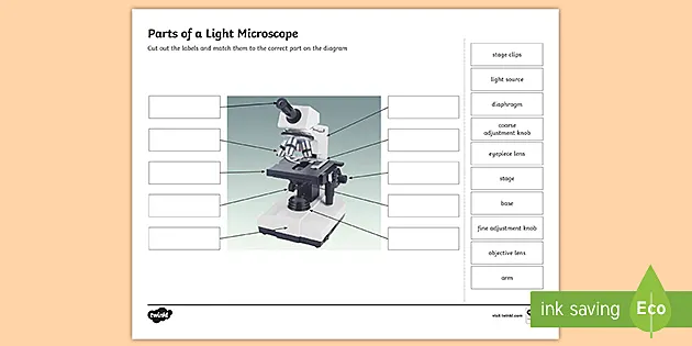
Parts of a Light Microscope Activity | Labeling Task
Simple Microscope - Parts, Functions, Diagram and Labelling They are the parts of the microscope that involved passing the light through the specimen and magnify its size. Parts of the optical parts are as follows: Mirror - A simple microscope has a plano-convex mirror and its primary function is to focus the surrounding light on the object being examined.
1.2: Microscopes - Biology LibreTexts
› cells › bactcellInteractive Bacteria Cell Model - CELLS alive Ribosomes: Ribosomes give the cytoplasm of bacteria a granular appearance in electron micrographs.Though smaller than the ribosomes in eukaryotic cells, these inclusions have a similar function in translating the genetic message in messenger RNA into the production of peptide sequences (proteins).

Parts of a microscope with functions and labeled diagram
Dissecting microscope (Stereoscopic or stereo microscope) Parts ... Labeled Diagram of Dissecting microscope (Stereoscopic and Stereo microscope) A typical stereo microscope has 6 major parts which are:. LED Illuminators: Typically dissecting microscopes have an LED light that that illuminates the exhibit that needs to be observed. Eyepiece: Each dissecting microscope has two eyepieces that is used to focus on the light has divergent pathways.

Microscope Parts and Functions - YouTube
ProSciTech WebLaboratory supplies and Lab equipment for Histology, Pathology, Light Microscopy, Electron Microscopy and specialist researchers.
The Compound Light Microscope Label the following parts on ...
Microscope Quiz: How Much You Know About Microscope Parts ... - ProProfs Welcome to the ultimate Microscope Quiz. This quiz will check how much do you know about Microscope Parts and Functions! The microscope has been used in science to understand elements, diseases, and cells. You must have used a microscope back in high school in the biology lab. Do you believe you understood how to use it? Take up the test and see.

Microscope World Blog: Biological Microscope Parts
Amazon.com: Magnifying Glasses 8X 15X 23X Magnifier LED … WebAbout this item . Double eye magnifying glasses magnifier loupe, with 2pcs adjustable LEDs to help it work in low-light conditions ; Left right double eye patches magnifierloupe with adjustable LED to help work in low-light conditions.Set of 2 magnifying glasses mounted on a one-size-fits-all eyeglass frame for easy hands-free operation.
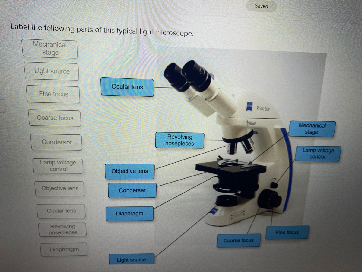
Answered: Saved Label the following parts of this… | bartleby
Microscope Parts, Function, & Labeled Diagram - slidingmotion Microscope Parts Labeled Diagram The principle of the Microscope gives you an exact reason to use it. It works on the 3 principles. Magnification Resolving Power Numerical Aperture. Parts of Microscope Head Base Arm Eyepiece Lens Eyepiece Tube Objective Lenses Nose Piece Adjustment Knobs Stage Aperture Microscopic Illuminator Condenser Lens

Parts of a Microscope - SmartSchool Systems
Confocal Microscope- Definition, Principle, Parts, Types, Labeled ... Parts of the Confocal Microscope The Confocal Laser Scanning Microscope is made up of a few components: Objective lens Out-of-focus plane In-focus plane Beam splitters Detector Confocal pinhole (aperture) Laser Oscillator Mirrors Types of Confocal Microscope
Microscope
Microscope Types (with labeled diagrams) and Functions Simple microscope labeled diagram Simple microscope functions It is used in industrial applications like: Watchmakers to assemble watches Cloth industry to count the number of threads or fibers in a cloth Jewelers to examine the finer parts of jewelry Miniature artists to examine and build their work Also used to inspect finer details on products
Parts of a Microscope with Their Functions – Microbe Online
Light Microscope- Definition, Principle, Types, Parts, Labeled Diagram ... Two focusing knobs i.e the fine adjustment knob and the coarse adjustment knob, found on the microscopes' arm, which can move the stage or the nosepiece to focus on the image. the sharpen the image clarity. It has a light illuminator or a mirror found at the base or on the microbes of the nosepiece.

Simple Microscope - Diagram (Parts labelled), Principle ...
Parts of the Microscope with Labeling (also Free Printouts) A microscope is one of the invaluable tools in the laboratory setting. It is used to observe things that cannot be seen by the naked eye. Table of Contents 1. Eyepiece 2. Body tube/Head 3. Turret/Nose piece 4. Objective lenses 5. Knobs (fine and coarse) 6. Stage and stage clips 7. Aperture 9. Condenser 10. Condenser focus knob 11. Iris diaphragm
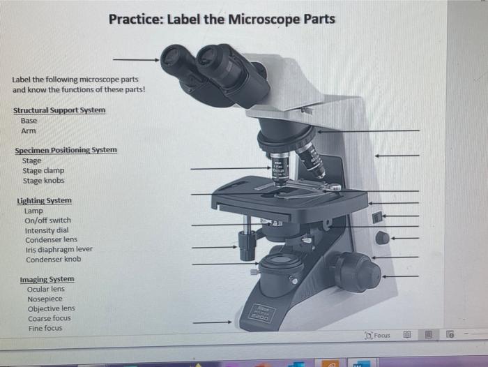
Solved Practice: Label the Microscope Parts Label the | Chegg.com
Compound Microscope - Diagram (Parts labelled), Principle and Uses What are the 13 parts of a microscope? 1. Eyepiece 2. Eyepiece Tube 3. Objective Lens 4. Stage 5. Stage Clips 6. Nosepiece 7. Fine and Coarse Focus knobs 8. Illuminator 9. Aperture 10. Iris Diaphragm 11. Condenser 12. Condenser Focus Knob 13. The Rack stop Q 5. What are the 11 parts of a compound microscope?
(159).jpg)
Microscope Quiz: How Much You Know About Microscope Parts And ...
Microscope: Parts Of A Microscope With Functions And Labeled Diagram. List down the 18 parts of a Microscope. Ocular Lens (Eye Piece) Diopter Adjustment Head Nose Piece Objective Lens Arm (Carrying Handle) Mechanical Stage Stage Clip Aperture Diaphragm Condenser Coarse Adjustment Fine Adjustment Illuminator (Light Source) Stage Controls Base Brightness Adjustment Light Switch

Compound Microscope Parts, Diagram Definition, Application ...
Sperm Under Microscope with Labeled Diagram - AnatomyLearner The sperm under a microscope with 40x, 100x, and 400x labeled diagrams might help you clear the basic concept. Categories Veterinary Histology Tags sperm under microscope Post navigation Binocular Microscope Anatomy - Parts and Functions with a Labeled Diagram
Parts of a Microscope | Microscope Parts and Functions | Labkafe
Phone Parts Wholesale Australia | iPhone Parts Wholesale WebCrazyparts is the leading Australian wholesale supplier of mobile phone parts, tablet parts and phone accessories, including iPhone Part, iPad Parts, Samsung Phone Parts, Mobile Phone Parts Repair and Replacement Parts etc. AU. AU. NZ. 1300 271 188 Mon-Fri 8:30AM-5:00PM 19-21 Euston Street Rydalmere, NSW 2116, Australia 1300 271 188 …

Label a Microscope Worksheet
Simple Squamous Epithelium under a Microscope with a Labeled Diagram ... In this portion, I will show you the simple squamous epithelium labeled diagrams from the different organs or parts, or structures of the animal's body. The typical example of the simple squamous epithelium will be found in the lung's alveoli, the parietal layer of the Bowman's capsule of the kidney, and the loop of Henle of kidney tubules.

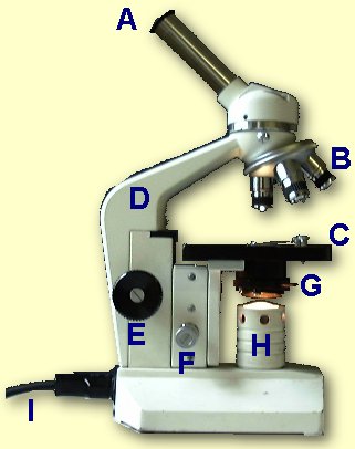
This is a common compound microscope. Label its parts from A ...
Light Microscope-Definition, Principle, Types, Parts, Labeled Diagram ... An alternative name for it is a compound light microscope. Parts of a bright-field microscope (compound light microscope) It is composed of: ... The light that was emitted is converted into an image that has been fluorochrome-labeled. The fluorescent microscope's working mechanism is based on the idea that by exposing the specimen to ultra ...
Modified Science Diagram; Label Parts of a Microscope; Special Education
Interactive Bacteria Cell Model - CELLS alive WebPeriplasmic Space: This cellular compartment is found only in those bacteria that have both an outer membrane and plasma membrane (e.g. Gram negative bacteria).In the space are enzymes and other proteins that help digest and move nutrients into the cell. Cell Wall: Composed of peptidoglycan (polysaccharides + protein), the cell wall maintains the …

Microscopy
rockyourhomeschool.net › microscope-worksheetsFree Microscope Worksheets for Simple Science Fun for Your ... Parts of a Microscope . The first worksheet labels the different parts of a microscope, including the base, slide holder, and condenser. If you have a microscope, compare and contrast this worksheet to it. Also, your kids can color this microscope diagram in and read the words to each part of the microscope.

Microscope labeling
Compound Microscope - Types, Parts, Diagram, Functions and Uses It comes with a wide body and base. Its distinct parts include a condenser, illumination, focus lock, mechanical stage, and a revolving nosepiece which can hold up to five objectives. It usually has a binocular head, which makes long-term observation easy. Image 22: An example of a research compound microscope.

Compound Microscope Parts
Microscopy- History, Classification, Terms, Diagram - The Biology Notes History of Microscope. In the 1 st Century AD, the Romans invented the glass and used them to magnify objects. In the early 14 th Century AD, eyeglasses were made by Italian spectacle makers. In 1590, two Dutch spectacle makers, Hans, and Zacharias Jansen created the first microscope. It was a simple tube with 2 lenses system and had 9X ...

Dissecting Stereo Microscope Parts and Functions
Barcode Labels and Tags | Zebra WebWith more than 400 stocked ZipShip paper and synthetic labels and tags – all ready to ship within 24 hours – Zebra has the right label and tag on hand for your application. From synthetic materials to basic paper solutions, custom to compliance requirements, hard-to-label surfaces to easy-to-remove labels, or tamper-evident to tear-proof, we have more …

Microscopy 101 | Parts of the Microscope | Microscope Central

Parts of a Compound Microscope and Their Functions
Parts of a Microscope Quiz
Parts of a microscope with functions and labeled diagram

label the parts of the compound microscope - Brainly.ph

Microscope Maintenance Tips | Science supplies, Multi step ...

Parts of the Microscope worksheet

Bellwork Why do scientists use Microscopes? - ppt download

Labeling a Microscope Free Worksheet Pack
Post a Comment for "40 microscope parts and labels"