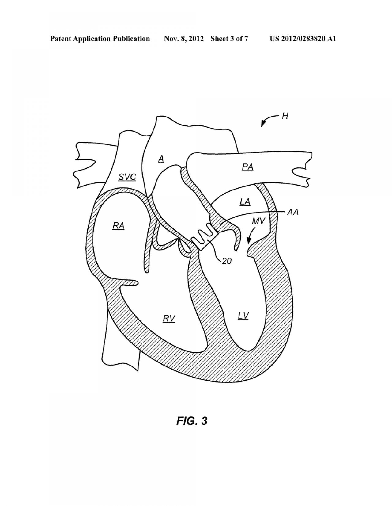
45 external structure of the heart with labels
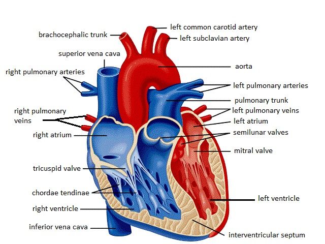
Heart Anatomy: Heart Dissection The letters indicated in the text refer to the labels on the picture. The anterior surface of the heart is characterized by the presence of the large arteries leaving the base of the heart, the pulmonary trunk (H) and the aorta (G). The pulmonary trunk is the vessel that divides to give rise to the two pulmonary arteries going to each lung. OpenStax AnatPhys fig.19.6 - Surface Anatomy of the Heart - English labels External Anatomy of the Heart. Inside the pericardium, the surface features of the heart are visible. English labels. From OpenStax book 'Anatomy and Physiology', fig. 19.6. Anatomical structures in item: Cor Truncus brachiocephalicus Vena cava superior Truncus pulmonalis Aorta ascendens Atrium dextrum Atrium sinistrum Ventriculus dexter
Heart - External Features - Anatomy QA Location of heart: Heart lies in the middle mediastinum. 1/3rd of the heart lies to the right and 2/3rd to the left of the midline. It lies opposite to T5 - T8 vertebrae in supine position & T6 - T9 vertebrae in erect position. Dimensions of heart: Base to apex-12cm; Transversely- 8-9cm; Anteroposteriorly- 6cm.

External structure of the heart with labels
How to Draw the Internal Structure of the Heart (with Pictures) To draw the internal structure of a human heart, follow the steps below. Part 1 Finding a Diagram 1 To find a good diagram, go to Google Images, and type in "The Internal Structure of the Human Heart". Find an image that displays the entire heart, and click on it to enlarge it. 2 Find a piece of paper and something to draw with. Lesson | The Heart - External Structure | Encounter Edu In this lesson students begin their exploration of the circulatory system, labelling a diagram of the external structures and identifying arteries and veins. They will go on to explain where blood enters and leaves the heart. Learning outcomes byjus.com › biology › human-heartHuman Heart - Anatomy, Functions and Facts about Heart The external structure of the heart has many blood vessels that form a network, with other major vessels emerging from within the structure. The blood vessels typically comprise the following: Veins supply deoxygenated blood to the heart via inferior and superior vena cava, and it eventually drains into the right atrium.
External structure of the heart with labels. quizlet.com › 574029087 › ch-19-circulatory-systemCh. 19 Circulatory System- heart Flashcards | Quizlet Correctly label the external anatomy of the anterior heart. Place the labels in order denoting the flow of blood through the pulmonary circuit beginning with the right atrium and ending in the left atrioventricular valve. The first and last structures are given. Right atrium 1. tricuspid valve 2. right ventricle 3. pulmonary valve techcrunch.com › gadgetsGadgets – TechCrunch Cultivated meat, grown in a bioreactor rather than out on the range, might be one of the big food trends of the decade. But it’s relying on tech built around multiplying yeast and bacteria cells Answered: Answer Label the dorsal external… | bartleby Label the dorsal external features of the frog's heart. Answer all labels on the space provided. 7. Transcribed Image Text: Instructions: 1. Label the dorsal and ventral external features of the frog's heart. Answer all labels on the space provided. 1. pulmonary vein 2. 3. PDF Anatomy of Heart Labeled and Unlabeled Images (a) Anterior view of the external heart C' 2019 Pearson Education. Aort'c arch Ligamentum arteriosum Left pulmonary artery Left pulmonary ve ns Auricle of left atrium Circumflex artery Left coronary artery (in atrioventricular sulcus) Great cardiac vein Left ventricle Anterior interventricular artery (in anterior interventricular sulcus) Apex
quizlet.com › 630625176 › chapter-19-the-heart-flashChapter 19: The Heart Flashcards | Quizlet •Allows heart to beat without friction, gives it room to expand and resists excessive expansion •Parietal pericardium-tough outer, fibrous layer of connective tissue-inner serous layer •Visceral pericardium (a.k.a. epicardium of heart wall)-serous lining of sac turns inward at base of heart to cover the heart surface Solved Heart Lab Worksheet ? Saved Help Save & Exit Submit - Chegg Anatomy and Physiology questions and answers. Heart Lab Worksheet ? Saved Help Save & Exit Submit Label the external features of the heart using the hints provided. 36 Pulmonary v 0.25 points Pulmonary trunk eBook Coronary sulcus Anterior interventricular sulcus Print References Base of heart Aorta Apex Right auricle Reset Zoom. External anterior heart labeling Quiz - PurposeGames.com This is an online quiz called External anterior heart labeling There is a printable worksheet available for download here so you can take the quiz with pen and paper. Your Skills & Rank Total Points 0 Get started! Today's Rank -- 0 Today 's Points One of us! Game Points 27 You need to get 100% to score the 27 points available Actions The structure of the heart - Structure and function of the heart ... The structure of the heart. If you clench your hand into a fist, this is approximately the same size as your heart. It is located in the middle of the chest and slightly towards the left.
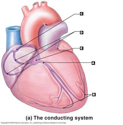
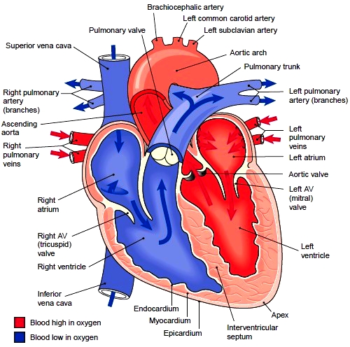
Internal Structure of the Heart | Contemporary Health Issues It is marked by the presence of four openings that allow blood to move from the atria into the ventricles and from the ventricles into the pulmonary trunk and aorta. Located in each of these openings between the atria and ventricles is a valve, a specialized structure that ensures one-way flow of blood. Heart Anatomy | Anatomy and Physiology - Lumen Learning The wall of the heart is composed of three layers of unequal thickness. From superficial to deep, these are the epicardium, the myocardium, and the endocardium. The outermost layer of the wall of the heart is also the innermost layer of the pericardium, the epicardium, or the visceral pericardium discussed earlier. Figure 6. Diagram of the human heart Images, Stock Photos & Vectors - Shutterstock Find Diagram of the human heart stock images in HD and millions of other royalty-free stock photos, illustrations and vectors in the Shutterstock collection. Thousands of new, high-quality pictures added every day. ... Anatomy. Healthcare and Medical. Diseases, Viruses, and Disorders. Icons and Graphics. heart. medicine. organ. human body. Chapter 22 Heart Flashcards | Quizlet Label the coronary arteries in an anterior view of the heart. Label the order that blood flows through in the heart, using the arrows as guides. Label the components of the heart wall. Label the components of the heart as seen from a posterior view. Label the major coronary veins. Label the components of the conduction system.
Unlabelled Diagram Of The Heart - Cliparts.co
Structure of the Heart | SEER Training The human heart is a four-chambered muscular organ, shaped and sized roughly like a man's closed fist with two-thirds of the mass to the left of midline. The heart is enclosed in a pericardial sac that is lined with the parietal layers of a serous membrane. The visceral layer of the serous membrane forms the epicardium. Layers of the Heart Wall

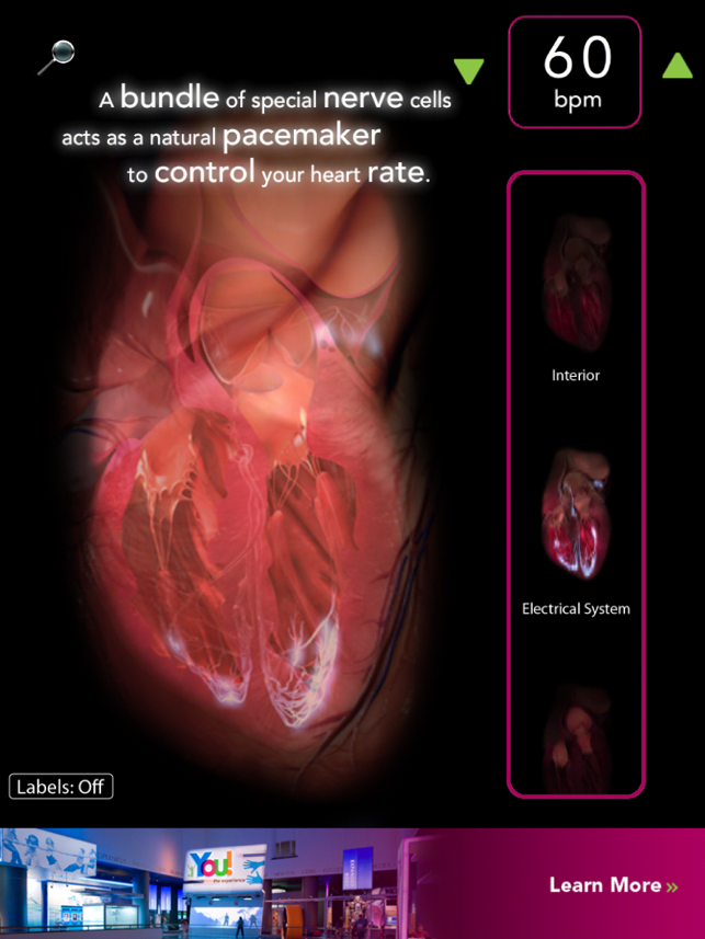
How Your Heart Works | Health Resources | Wellness Library ...
Correctly label the following external anatomy of the anterior heart ... Saved Correctly label the following external anatomy of the anterior heart. 18 Apex of heart 0.37 points Ascending aorta Skipped Ligamentum arteriosum Left pulmonary veins References Pulmonary trunk Anterior interventricular sulous Left auricle Left...
Virtual Heart on the App Store
Label The Heart Worksheet Label The Heart Worksheet. This is a simple and free human heart anatomy chart for kids. Left atrium right atrium left ventricle right ventricle aorta pulmonary veins pulmonary artery superior vena cava inferior vena cava bicuspid valve tricuspid valve aortic valve pulmonary valve. Featuring six of the basic parts of the heart each numbered and ...

Biology Diagrams,Images,Pictures of Human anatomy and physiology ...
The Anatomy of the Heart, Its Structures, and Functions Updated on April 05, 2020. The heart is the organ that helps supply blood and oxygen to all parts of the body. It is divided by a partition (or septum) into two halves. The halves are, in turn, divided into four chambers. The heart is situated within the chest cavity and surrounded by a fluid-filled sac called the pericardium.
iGCSE Biology - Gross Structure Of The Heart - BioChem Tuition
Label the external anatomy of the heart. Reset The McGraw-Hill ... Label the external anatomy of the heart. Reset The McGraw-Hill Companies, Inc. Photos and Dissections by Christine Eckel Ascending aorta Left pulmonary vein Pulmonary trunk Branch of the right pulmonary artery Right atrium Left ventricle Right ventricle (13a 2 -6a 5 -2a) - (-10a 2 -11a 5 +9a) 6. (7-13x 3 -11x) (2x 3 +8x - 4x 5 )
Chapter 20 The heart Flashcards | Easy Notecards
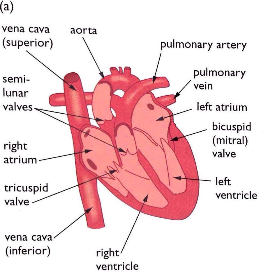
Describe the internal structure of human heart. - Toppr Solution Verified by Toppr The heart is divided into a right and left side by the septum. The heart has four chambers, two relatively small upper chambers called atria and two larger lower chambers called ventricles. The walls of the ventricles are relatively thicker than atrial walls.
Free Blank Heart Diagram, Download Free Blank Heart ...
Heart Diagram with Labels and Detailed Explanation - BYJUS Diagram of Heart. The human heart is the most crucial organ of the human body. It pumps blood from the heart to different parts of the body and back to the heart. The most common heart attack symptoms or warning signs are chest pain, breathlessness, nausea, sweating etc. The diagram of heart is beneficial for Class 10 and 12 and is frequently ...

Science&Life: Heart Dissection
art-labeling activity: external anatomy of the sheep heart Imagine the heart in the body of a person facing you. Internal anatomy of the heart 1 of 2 Part A Drag the labels to identify structural components of the heart. Gross anatomy of the stomach. This image shows an external view of a preserved sheep heart. Right atrium receives su. An unregistered player played the game 1 hour ago. The heart is ...

Biology 156
Heart Anatomy: Labeled Diagram, Structures, Function, and Blood Flow Let's begin with the chambers of the heart. There are 4 chambers, labeled 1-4 on the diagram below. To help simplify things, we can convert the heart into a square. We will then divide that square into 4 different boxes which will represent the 4 chambers of the heart.

Aqua Fanatic: Crayfish Anatomy
› articles › flat-vs-deep-hierarchyFlat vs. Deep Website Hierarchies - Nielsen Norman Group Nov 10, 2013 · Left: a flat site hierarchy, with few vertical levels. Right: a deep site hierarchy has the same information organized into more sublevels. Both of these site hierarchies start at the top with a single homepage, but the information below that page is organized quite differently: the website on the left has 8 major categories, but the site on the right has only 4.

1000+ images about School on Pinterest | Heart diagram ...
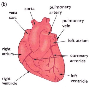
pmt.physicsandmathstutor.com › download › BiologyPractical notes - SP 2.3c Dissection of a Mammalian Heart ... The mammalian heart is a muscular pump that pushes blood around the body. It consists of four chambers and associated blood vessels . The left and right side of the heart is separated by a muscular wall, the septum . Recall the structure of the heart in the diagram below:
Heart. Structure of the Heart. Divisions of the Heart
en.wikipedia.org › wiki › The_TenorsThe Tenors - Wikipedia The Tenors (formerly known as The Canadian Tenors) are a vocal group consisting of Victor Micallef, Fraser Walters, and Clifton Murray.They perform operatic pop music that is a mixture of classical and pop, featuring songs such as "The Prayer", Panis angelicus, and Leonard Cohen's Hallelujah.
iGCSE Biology - Gross Structure Of The Heart - BioChem Tuition
External Heart Anatomy Quiz - PurposeGames.com This is an online quiz called External Heart Anatomy. There is a printable worksheet available for download here so you can take the quiz with pen and paper. Your Skills & Rank. Total Points. 0. Get started! Today's Rank--0. Today 's Points. One of us! Game Points. 27. You need to get 100% to score the 27 points available.

Heart Anatomy Using Models
Solved -labeling Activity: External Anatomy of the Sheep | Chegg.com Anatomy and Physiology. Anatomy and Physiology questions and answers. -labeling Activity: External Anatomy of the Sheep Heart Part A Drag the labels to the appropriate location in the figure. Reset Help Lolt ventric Pulmonary trunk Lolt atrium Lohtaude Right trum Posterior Interventricular sules = Pulmonary veins Art Right vorticle Anterior ...

u414adad: heart diagram without labels
Structure Of The Heart | A-Level Biology Revision Notes The heart is a hollow muscular organ that lies in the middle of the chest cavity. It is enclosed in the pericardium, which protects the heart and facilitates its pumping action. The heart is divided into four chambers: The two atria (auricles): these are the upper two chambers. They have thin walls which receive blood from veins.
Post a Comment for "45 external structure of the heart with labels"